Today's article comes from the IEEE Access journal. The authors are Almalki et al., from Temple University, in Philadelphia. In this paper they introduce a new system (called MDEP) for dental cavity detection.
DOI: 10.1109/ACCESS.2025.3606811
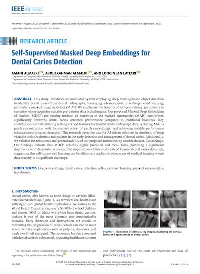
Got a toothache? Well, you're not alone. Dental cavities are one of the most common diseases on the planet. More than half of school-children and nearly every adult has experienced them at some point. That means that cavity-detection is one of the most frequently performed medical diagnoses in the world. But despite all that, the process of detecting and identifying cavities (or "caries") is actually still quite prone to human error.
Side note: Dental caries is the scientific term for the disease process that gradually breaks down tooth enamel and dentin. A cavity is the visible hole or structural damage that results once the caries process has progressed far enough. In practice, the two words often get used interchangeably.
Traditionally, caries detection relies heavily on two things: visual-tactile examinations and radiography. Both methods have significant limitations.
These approaches are fine in many cases, but there are instances where they result in delayed diagnosis and treatment, increasing patient inconvenience and overall costs.
That's where this paper comes in. The authors have built an automated system that detects dental caries from radiographs using a technique called Masked Deep Embeddings of Patches, or MDEP. On today's episode we're going to explore how MDEP works, how it scales, and whether or not it's actually better than having a dentist look at your x-rays. Let's dive in.
In medical imaging, labeled datasets are often limited. You can't just scrape millions of dental X-rays from the internet like you could with cat photos. And once you have the images, they need to be annotated by a qualified professional. This is time-consuming and costly, and creates a problem for traditional deep learning approaches (that require vast amounts of labeled data to perform well). The solution lies in self-supervised learning, specifically a technique called masked image modeling. Picture it like this: you're teaching a model to recognize dental features by showing them X-ray images with certain patches covered up, then asking them to predict what should be in those hidden areas. By learning to fill in the blanks, the model develops a deep understanding of dental anatomy and pathology without needing explicit labels for every single feature.
This particular implementation is built on an existing technique called Masked Autoencoders, but with a twist. Instead of reconstructing the pixel values of masked patches (which can be noisy and less meaningful), their MDEP method focuses on reconstructing the deep embeddings of those patches. As a reminder: embeddings are not pixels, think of them as rich, mathematical representations that capture the essential characteristics of image regions. This approach leads to more semantically meaningful reconstructions than if you used the pixels themselves.
First, they divide input radiograph images into non-overlapping patches. These patches are then embedded using a multi-layer perceptron to create patch embeddings. During the pre-training phase, a subset of these patches is randomly masked. The masked embeddings are replaced with a shared learnable mask embedding, while positional embeddings are added to retain spatial information.
The encoder, which is based on a Vision Transformer architecture, processes only the visible patches. This is computationally efficient because it doesn't waste processing power on masked regions during training. In case you're unfamiliar, Vision Transformers are quite a shift from traditional CNNs. Instead of using convolution operations that analyze local neighborhoods of pixels, Vision Transformers treat images as sequences of patches and apply an attention mechanism to them. That attention mechanism is what makes Vision Transformers particularly powerful. Traditional convolutional networks have a limited receptive field, meaning each neuron can only see a small portion of the image at any given layer. Vision Transformers, through that attention mechanism, can relate any patch to any other patch in the image, regardless of their spatial distance. This global context is exactly what's needed for dental diagnosis, where the relationship between different anatomical structures can provide important diagnostic information.
Back in pre-training a lightweight decoder was actually appended to the encoder specifically for predicting the latent representations of masked patches. So the decoder receives both the encoded visible patch embeddings and placeholder mask tokens with positional information. It then attempts to reconstruct the original embeddings corresponding to the masked patches. Rather than using mean squared error in pixel space as traditional autoencoders do, this system computes what's called L1 loss between the original and predicted embeddings of masked patches. L1 loss measures the absolute difference between predicted and actual values, (rather than the squared difference used in mean squared error). This makes the loss function more robust to outliers and can lead to sharper, more accurate embeddings.
After pre-training, the decoder is discarded entirely and the encoder serves as the backbone for a Mask R-CNN with Feature Pyramid Network. More on both of those is a moment. The pre-trained Vision Transformer weights initialize the encoder for the detection task. Features extracted by the Vision Transformer backbone are passed to both the neck and the detection head to facilitate bounding box regression and classification.
The Feature Pyramid Network helps build high-level semantic feature maps at different scales. Traditional object detection methods struggle with objects of varying sizes because they typically operate at a single scale. Feature Pyramid Networks solve this by creating a pyramid of features at multiple resolutions, allowing the model to detect both small and large cavities effectively. The Mask R-CNN provides pixel-level segmentation information alongside bounding box detection. While bounding boxes tell you where a cavity might be located, segmentation masks tell you exactly which pixels belong to the cavity. This precision is valuable because it allows for more accurate measurement of cavity extent and better treatment planning.
Training has two stages. During the self-supervised pre-training phase, the system learns general representations of dental anatomy without requiring labeled examples of cavities. This is where the masking strategy comes into play. The model learns to understand dental structures by being forced to predict missing pieces of radiographs based on the visible context. For the downstream fine-tuning phase, the system adapts its learned representations to the specific task of cavity detection using labeled data. But, (and this is the whole point), since the model has already learned rich representations during pre-training, it requires far less labeled data to achieve high performance on detection. The entire network effectively undergoes fine-tuning to perform the detection task. The pre-trained weights serving as strong initialization that allows faster convergence and better performance with limited training data. This, in effect, is transfer learning.
The authors validated this approach on two datasets:
For evaluation they used standard object detection metrics (precision, recall, and Average Precision, etc). The results demonstrate substantial improvements over the baselines. On both of the datasets MDEP significantly outperformed the baselines. And the fact that it works so well on both types of images bodes well for its generalizability.
While the numbers are impressive, real-world deployment faces practical challenges. From technical issues like class imbalance in patient populations, to more logistical concerns like integration with existing clinical workflows and regulatory approval. That being said, those are probably more speedbumps than roadblocks. If the results they achieved here are replicable and generalizable, it should only be a matter of time before we start to see people trying this out in the wild.
If you want to dive deeper into the derivations behind the MDEP loss function, or explore the ablation studies, I'd highly recommend that you download the paper. The authors also include a breakdown of the architectural modifications they made to the Vision Transformer, and this will be very useful should you plan on building this kind of system yourself.